Sunday, April 9, 2017

The Electrospinner pt 2

Ah yes, part 2. 

Even though my last post was quite extensive in detail, there is still more to talk about! But before I get into that, I have to introduce two new faces: Max and Tatum (both undergrad students). Now, unlike my previous posts, I cannot go into extensive details due many of the methods and details being proprietary.

As I mentioned last time, the electrospinner creates scaffolds. These scaffolds can then be placed in a wound in order to accelerate healing. But, there's one problem: pathogens. While the wound heals, the site needs to be protected from deadly bacteria (pathogens) that could potentially impair wound healing. So the question is how to protect the wound? 

This leads us to Max and Tatum. One of the ideas that they are exploring is the combination of scaffolds with an antimicrobial therapeutic. So what exactly does this mean? In order for this to make sense, you have to remember how electrospinning works: we use a gelatin to create the scaffolds. The gelatin mixture is pumped from the syringe and sprayed onto the target, ultimately making a scaffold. Now, since we can modify what we put in the syringe, we can modify the properties of the scaffold. In this case, if we add an antimicrobial therapeutic to the gelatin in the syringe, we can create a scaffold resistant to harmful bacteria. 

Now that's pretty cool. 

At the moment, we are still experimenting with how much of the therapeutic we should add to the scaffold. Too much, and it might be a waste. Too little, and bacteria might still grow. So this is why we spin multiple scaffolds, each with different amounts of added therapeutic. But here's the catch, each scaffold takes an hour to spin. Let's just say, lots of music was listened to while waiting...

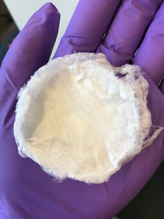
The end result:


As you can see, nothing looks out of the ordinary; it's just another scaffold. But inside, it has a secret weapon: antimicrobial therapeutics. Pathogens beware! 

Now that the scaffolds are all set and done, we have to test them. Each scaffold will be placed into a dish with growing bacteria. Depending on the results, we may be one step closer to creating a pathogen-resistant scaffold.

-Tudor
Wednesday, March 22, 2017

The Electrospinner

What if every time you went for a drive, you had to build the road in front of you?

Not only would that suck, but it would take forever to get anywhere. But, this is exactly how wound healing works. Cells have to lay down the "road" in order to grow. Cells can not automatically start growing in a wound. In fact, they require a structure (or matrix) to grow and move on. A release of matrix proteins creates this matrix structure, but this only happens one fragment at a time since the proteins are released as the cells move. This is then continued until the wound is closed. As is the case with the driving analogy, this process is slow. Very slow.

But what if this process could somehow be accelerated? What if the matrix was already in place before the cells started moving? Since I'm writing about this, you've probably realized that there must be a way to do just that. And yes, there is! With the help of an electrospinner, a scaffold can be created. From there, the scaffold can be inserted inside a wound, which will give the cells an open highway to start moving on. So now you're probably wondering, what's an electrospinner? To put it simply, it's a fancy machine that creates scaffolds:*
Electrospinner setup. A) syringe pump. B) Electrospinning cage for maintenance of humidity and electrical current. C) syringe needle for distribution of gelatin fibers. D) target. E) power supply.
But of course, the electrospinner can not make a scaffold from out of thin air! As the photo caption above hints, gelatin is used to create these scaffolds. Currently, two different gelatin's are being used: bovine and porcine. Bovine, which comes from cows and porcine which comes from pigs. The cool thing about gelatin is that it contains absolutely zero cells, which means that it will be easily accepted into our body regardless of the source. 

Why does this matter? 

Because, anything non-biological is considered really foreign to the body. So the best option is to use something biological. And sure, cells are biological, but if they don't belong to us, they're pretty foreign. So here's gelatin to save the day! At the moment, we have not determined which gelatin (bovine or porcine) is better for creating the scaffolds, so both are being used. 

Now, the actual creation (or spinning) of the scaffold takes about an hour. This means, an hour of waiting and watching. It would be nice to leave the machine to do it's thing, but every now and then a falling fiber has to be moved or cleaned off the syringe needle. It may be a bit hard tell, but in the photo below the fibers are coming out of needle and landing on the target (a circular piece of aluminum foil):*


  Once this is all set and done, we have ourselves a scaffold!* 


I can now say I've made a highway. A highway for cells.

-Tudor


*A big thanks to Martha and Tatum for the photos!

Wednesday, March 8, 2017

Scratch Assay pt. 2

And we're back! This time, with an actual experiment. 

Since the cells from last time didn't grow to full density, new cells had to be grown. This took about a week. Now, the experimental set-up remains the same as previously stated: scratch assays will be used to observe the effects of arsenic and estrogen in a wound. Theoretically, the arsenic contaminated wounds should take longer to close than the control, while the estrogen treated wounds should close around the same time as the control. 

I have to pause here and give credit to Bronson and Oscar. I would love to say that I performed this experiment, but let's be real here, that did not happen. Together, they did the work while I sat back and observed (but don't worry, I took notes). Now just as before, we had to check the growth density of our cells. Fortunately, nothing was stopping us this time! From here on, the actual step by step process of what happened is not worth noting, but here's a summary: The cells were moved from their original container to a "plate" consisting of 12 different wells. Since the plate can be divided into 3 rows, each row represented a different variable (control, arsenic contamination, and estrogen treatment of arsenic contaminated wounds). Once all of this was taken care of, Bronson scratched the cells with a pipette tip. From there, the wells were placed in the incubator to grow. And then we waited.

Every 4 hours, over a 20 hour time period, photos where taken of the wells. The results:


So as expected, the arsenic contamination slowed the wound closure. But, as shown above, estrogen treatment almost reversed the contamination's obstruction to the healing process. Now that's pretty cool.

-Tudor

Edit: I almost forgot...all of this started at 6 in the morning. Bronson...

Friday, February 17, 2017

Scratch Assay

Finally, an experiment!

But before the fun begins, I have to introduce two new faces: Bronson (PhD Student) and Oscar (Undergrad Student). Together, they will be conducting the experiment while I observe. 

But here's the catch.... I had to wake up at 4:45 am. Yes, am. In order to view said experiment, I had to be on NAU's campus at 6:00 am. Now, there's no particular reason that the experiment had to be done this early, but according to Bronson, he's more of a morning person when it comes to doing research. Ugh, I am not a morning person no matter the cause. But it had to be done. For the greater good. For science! (okay, maybe a bit too dramatic)

Anyways, once on campus, I couldn't just stroll into the lab as I had imagined. Nope, the building's exterior doors were locked. I mean, sure, makes sense. It's not like anyone was expecting visitors at 6 in the morning. But, when you're half asleep and freezing, these thoughts don't go through your mind (at least, not mine). Long story short, Oscar managed to save me from turning into a human popsicle. 

Once in the lab, we couldn’t immediately start the experiment. Since we were creating scratch assays (hence the title), cells had to be grown a full week in advance to use today. So, the cell growth density for each sample had to be checked. This is important because, if the cells don’t grow to full density (pretty much cell next to cell) the test is scrapped, since the sample will not mock human skin. As it turned out, both samples were not fully grown. 

Now, why this happened, is still a big unknown. But regardless, the day wasn’t completely wasted. While we were sitting around wondering what could've gone wrong, we also discussed the experimental set-up. Here's what I learned: In general, scratch assays are effective for mimicking a wound in vitro. In this case, the assays will also be used to observe the effects of arsenic and estrogen in a wound. In theory, the arsenic-contaminated wounds should take longer to close than the control. Continuing this thought, the wound that has been treated with estrogen should hopefully close faster than the control. Now, the big question is, what happens if you treat the arsenic-contaminated wound with estrogen? Could the contamination process somehow be reversed? Only experimentation will tell.

Science... it can be a pain sometimes

-Tudor

Sunday, February 12, 2017

The Works

Before we can dive into the science of artificial* wound healing, we have to understand how natural healing occurs. 

Our skin serves as the first protective barrier to outside pathogens. But once the skin is penetrated and damaged, three consecutive stages will take place: inflammation, proliferation, and remodeling. Local inflammation serves an important role since it becomes a sort of "activator" for further responses. One such response is the release of chemical signals, which will ultimately help deliver clotting agents and phagocytes to the wound. If you're scratching your head wondering what these are, don't worry! I'm here to help.

Clotting agents will, simply enough, clot. This will prevent the loss of too much blood, but we're not done yet. Foreign material that flooded the wound, whether this be specks of dust or pathogens, has to be taken care of. Luckily for us, phagocytes love to eat material, but only foreign material (which is good, or else they would be engulfing our bacteria as well). From here on, antibodies become activated and blah blah blah... Admittedly, I am going to skip over quite a few steps here, but, they're not essential to understanding wound healing. In fact, the remaining immune responses deal with cleaning up the mess: destroying the antigens (foreign material) and then creating immunity to the antigens.

At this point, we still have a gash in our skin. So, proliferation takes place: the cells surrounding the wound begin to reproduce rapidly and move across the wound. Even once the wound is closed, the cells are still at work. During the remodeling phase, the dermal tissues continue to reproduce in order to increase strength and support. 

Now, all these steps were somewhat simplified. But regardless, the scope of artificial wound healing is to improve on these existing steps. This can be in the form of accelerating the entire process, or even creating new skin for the scarring that remains. 

-Tudor


*in the sense that the healing has to be promoted by some outside factors

Sunday, February 5, 2017

Introduction!

Hello there!


My name is Tudor. Yes, like the English Dynasty (and no, I was not named after them).

For the next twelve weeks I will be working on my Senior Research Project (SRP), which I have dedicated to wound healing. I will be focusing this project and blog on the latest research behind this issue. Now, wound healing in itself is a very broad topic, which leaves a lot of room to explore. But the truth of the matter is that key problems, such as sustainability and efficiency, lead the focus while also opening new doors to the unknown. For example, skin grafts require a donor. This means that the skin, in lack of a better phrase, has been used. Not only this, but finding a donor is also a problem. As is usual for any progress, someone asked "isn't there a better way?" And voilĂ , we have a need for research!

This research will be taking place at Northern Arizona University and at Northern Arizona Center for Entrepreneurship and Technology (NACET). I am super grateful to be working alongside Dr. Robert Kellar and his students. I am also grateful for the guidance and support that my on campus advisors, Ms. Alicia Vaughan and Ms. Lisa McDonough, will provide me.

Now let's science the heck out of this!